Four Decades of Innovation in Biological Electron Microscopy
- Pioneered electron microscopy of live cells, and electron diffraction of protein crystals, in liquid water
- Invention of the “single-particle” method for 3-D reconstruction of non-periodic specimens from random EM projections
- First 3-D reconstructions of cellular structures (mitochondria, axonemes, kinetochores) by EM tomography
- First cryo-EM structures of bacterial, mammalian mitochondrial, and chloroplast ribosomes
- First 3-D EM reconstructions using EM stereoscopic contouring within a single specimen
- Pioneered use of microcapillary pipettes to image specimens (e.g. patch-clamped membranes) and compute tomograms over the full 360° tilt range
- First EM tomograms incorporating projections from a specimen tilted around two orthogonal axes
- First 3D reconstructions of frozen tissue obtained by EM tomography
- First North American use of EM phase-plates to image frozen specimens
- First EM tomograms from frozen cells milled with a focused ion beam
- First 4-D EM reconstruction of macromolecular assemblies (ribosomes), utilizing a microfluidic mixer-sprayer to freeze macromolecules in time-resolved states

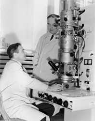
Helmut Ruska, MDM (standing) was a pathologist at Wadsworth in the 1950s,and brother of Ernst Ruska who won the Nobel prize for his invention of the EM. Seated at the Siemens Elmiskop I TEM is Dr. George Edwards, then Director of Micromorphology.